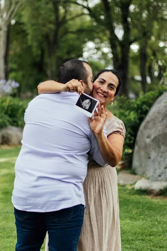

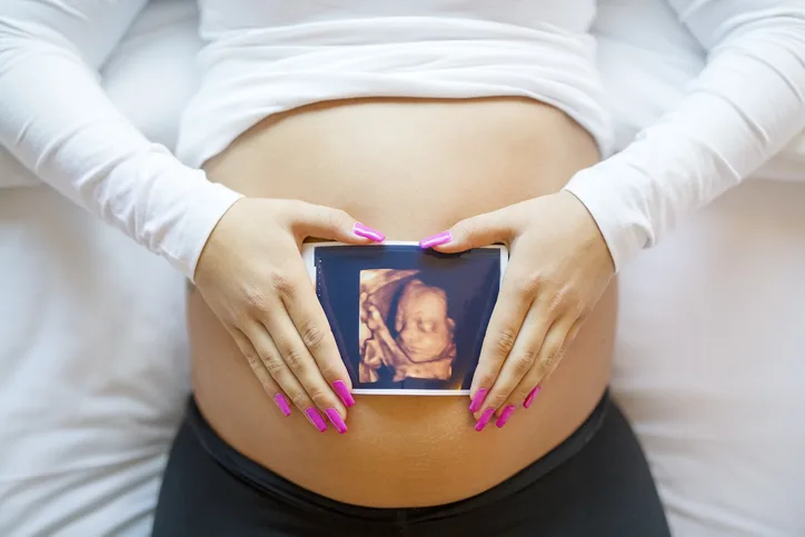
Obstetric Ultrasound
Many parents are excited to know when their baby can be seen on ultrasound. Two methods of obstetrical ultrasound are performed. A transvaginal ultrasound, usually performed in the first trimester, requires the bladder to be empty and involves placing a narrow transducer into the vagina for better visualization of the early pregnancy. An abdominal ultrasound, which is usually performed in the second and third trimesters, requires a full bladder and involves placing the transducer over the abdomen and is used for visualizing the uterus and the fetus. You will be instructed on how to prepare for your exam at the time the appointment is scheduled.
We are able to gain information such as the estimated size of the fetus, rate of growth of the fetus, determine the number of fetuses, fetal position, and detect some birth defects.
Our information brochure "Meet Your Baby Early" provides information on the portrait options available to you.
To assist you in planning for your ultrasound, we do ask that you review our Guidelines for the Sonography Room prior to your visit
3D and 4D Ultrasound
Seven Oaks Women's Center is excited to offer 3D and 4D ultrasounds for our expecting mothers! With advancements in today's technology, you can view your baby with more details than ever before. Learn more about 3D and 4D ultrasounds
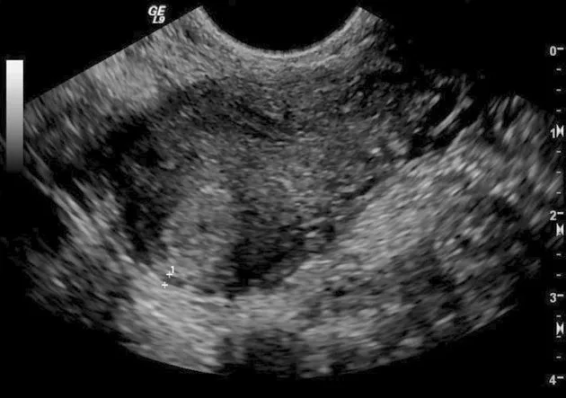
Gynecologic Ultrasound
With a gynecologic ultrasound, we are able to see and evaluate the pelvic anatomy and better visualize abnormalities such as uterine fibroids, endometrial polyps, and ovarian cysts.
Most gynecologic ultrasounds are best performed transvaginally with a narrow transducer inserted into the vagina. At times, an abdominal ultrasound may be used.
Also, 3D imaging may be performed to better visualize anatomy such as uterine fibroids.
Sonohysterogram
A sonohysterogram is a specialized ultrasound that allows your physician to visualize the endometrial cavity, or lining of the uterus, in more detail than a standard transvaginal ultrasound. This may be performed to detect the presence of a uterine polyp or fibroid that is distorting the endometrial cavity, look for scar tissue inside the uterus, or abnormal uterine shape. This exam is performed with both your physician and the ultrasonographer in the room. The physician will insert a small flexible tube into the cervix using a speculum. Fluid is pushed into the uterus through the tube while the ultrasonographer is using the abdominal ultrasound to capture images of the uterus.

Locations
Our Locations
We proudly serve patients needing gynecology services, obstetrics and overall women's health in the following areas:
Alamo Heights, Olmos Park, Castle Hills, Medical Center, Hill Country, Stone Oak, Hollywood Park, Downtown, Bulverde, Spring Branch, Leon Springs, Boerne, Alamo Ranch And Many Other Neighborhoods In The Greater San Antonio.
Medical Center
7707 Ewing Halsell,
Suite 213
San Antonio, TX 78229
Monday - Thursday:
8:30 a.m. - 5:00 p.m.
Friday:
8:30 a.m. - Noon
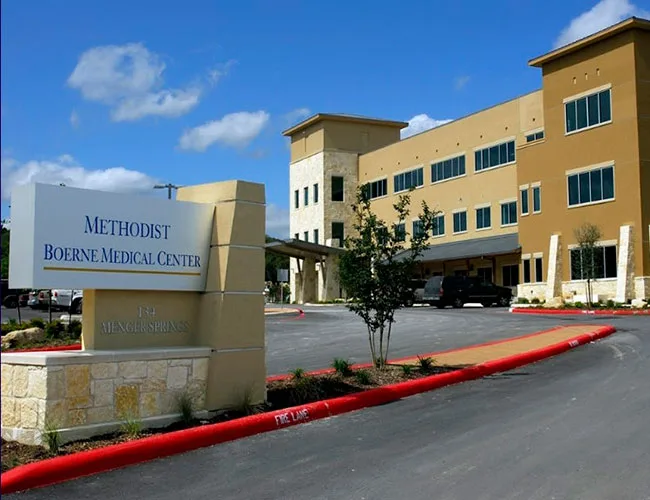
Boerne
134 Menger Springs,
Suite 1280
Boerne, TX 78006
Monday - Thursday:
8:30 a.m. - 5:00 p.m.
Friday:
8:30 a.m. - Noon